Gdańsk
Gdańsk, Plac Heweliusza. Osada. nr inw. Nr 343/97, wykop 29, warstwa 833.
Chronologia: XI–XIII w.
Elementy kończyny miednicy od pojedynczego osobnika w wieku dorosłym, o wysokości w kłębie 125,6-127,4 cm.
Literatura: Makowiecki 1998a.
|
Anatomical element |
NISP |
Descriptions and biometrical data |
|
Femur |
1 |
right |
|
Tibia |
1 |
right; GL = 323.5; SD = 33.4; Bd = 69; WH = 127.4 cm |
|
Third metatarsal bone |
1 |
right; GL = 240.5; Ll = 232.2; Bp = 41.9; SD = 41.9; Bd = 46; WH = 125.6 cm |
Gdańsk, 1; Grodzka St. 10/11, Grodzisko. nr inw. Nr 207, warstwa 430.
Chronologia: koniec XI w.
Elementy anatomiczne ze szkieletów kilku osobników (prawdopodobnie 4); każdy w wieku około 5–7 i 7–8 lat, dwóch w wieku 11–14 lat i jeden w wieku 14–16 lat, w tym dwóch mężczyzn.
Literatura: Makowiecka i Makowiecki 2008a; Jarzęcka-Stąporek i in. 2008.
|
Anatomical element |
NISP |
Descriptions and biometrical data |
|
Skull |
1 |
fragment of the occiput |
|
Skull |
1 |
incomplete – the entire braincase with the basal and visceral parts preserved, however, with the loss of the left orbital bone; nuchal crest slightly convex; left asymmetry; obtuse angle, nuchal ligament retracted, flat, but quite porous-rough; A = 1 B = 2 (27mm); individual about 7 years old; negatives of teeth visible on the right condyle, left condyle and condylar process knocked off – chipped off (blunt, heavy instrument) |
|
Mandible |
1 |
left, whole and part of the right, including tooth P2, wear of the crown P2 as a result of the bit, male aged 11-12 (based on incisors); incisors arranged almost horizontally |
|
Mandible |
1 |
left, strong abrasion of P2, male aged 14-16 (based on P3 measurement) |
|
Mandible |
1 |
left, strong abrasion of P2 due to the bit, age 12-14 |
|
Mandible |
1 |
right, small fragment, age 11-13 (based on P3 measurement) |
|
Axis |
1 |
|
|
Cervical vertebrae |
2 |
+-; circa 5–7 years |
|
Thoracic vertebrae |
6 |
+- ; circa 5–7 years |
|
Thoracic vertebrae |
1 |
process |
|
Lumbar vertebrae |
1 |
dog tooth marks |
|
Sacrum |
1 |
traces of gnawing by a dog in the last segment |
|
Sacrum |
1 |
|
|
Ribs |
16 |
|
|
Third metacarpal bone |
1 |
left; GL = 199.8; Bp = 45.5; SD = 29.4; Bd = 44.3; WH = 125.1 |
|
Pelvis |
1 |
right, left, traces of dog gnawing on the hip and ischial bone, very short individual – the size of a pony |
|
Pelvis |
1 |
left, preserved ilium with acetabulum, traces of dog teeth on the ilium; LA = 53.6 |
|
Pelvis |
1 |
right ilium, traces of gnawing by a dog |
|
Femur |
1 |
right, preserved proximal epiphysis and almost the entire diaphysis, traces of dog gnawing, traces of surface weathering from the caudal side |
|
Talus |
1 |
|
|
Third metatarsal bone |
1 |
right; Bd = 50 |
|
∑ |
42 |
|
Gdańsk, 1; Grodzisko. nr inw. Nr 204, warstwa 740.
Chronologia: 2. połowa XI w.; 14C: 955 ± 30 BP, nr laboratorium: Poz-155386; prawdopodobieństwo 68,20%: 1035AD (11,2%) 1049AD; 1081 r. (57,1%) 1152 r.; Prawdopodobieństwo 95,40%: 1027AD (95,4%) 1160AD.
14C: 900 ± 30 BP, nr laboratorium: Poz-155385; prawdopodobieństwo 68,20%: 1050AD (23,7%) 1081AD; 1153 r. (44,6%) 1212 r.; prawdopodobieństwo 95,40%: 1042AD (34,9%) 1108AD; 1116 r. (60,5%) 1219 r. n.e.
Elementy szkieletu co najmniej trzech osobników, w tym dwóch samców (czaszki), w wieku 7–8 lat i 12–13 lat; Wysokość w kłębie waha się od 122,9 do 129,6 cm; 135,6-139,9 cm i 142 cm. Dodatkowo w palcach osobnik bardzo krótki, około 115,1 cm.
Literatura: Makowiecka i Makowiecki 2008a; Jarzęcka-Stąporek i in. 2008 i ten projekt.
|
Anatomical element |
NISP |
Descriptions and biometrical data |
|
Skull |
1 |
male 12-13 years old; whole skull – very well preserved; smooth surface, light yellow; forehead profile (on the frontal bone) slightly concave; on the left nasal bone, in the area of the nasofrontal suture, diagonal scratches were noted (very delicate – barely visible to the naked eye, except for one distinct, negative, light knife with a very narrow blade); furthermore, two short, deeper (lenticular) negatives of the blade visible on the right nasal bone, located at the level of the junction with the nasal process of the incisive bone; on both nasal processes of the incisive bone visible negatives from the teeth of a predator (dog?) as shallow and wide, dark (brown) – the same in the left eye socket; on the nasal process of the right nasal bone, modifications of the original morphology are visible in the form of a punctate, shallow depression with a diagonal course, slight folds and thickening of the bone around it (the effect of injury, short-term inflammation); straight nuchal crest, left asymmetry, right angle; external occipital protuberance – convex with a visible spongy structure, a strongly emphasised process above it; 14C: Poz-155386; B-P = 477.5; WH = 136.8 cm; A-P = 518.2; WH = 139.9 cm |
|
Mandible |
1 |
left and right, 30 fragments with the left dental arch, traces of abrasion on the edge of P2 and the crown – after the bit; individual 11-12 years old |
|
Atlas and axis |
2 |
both fully preserved |
|
Cervical vertebrae |
1 |
last, preserved whole, individual over 10 years old (++), no signs of weathering |
|
Cervical vertebrae |
5 |
Whole, individual over 10 years old (++); the same individual |
|
Thoracic vertebrae |
10 |
over 10 years (++) three vertebrae with osteophytes |
|
Thoracic vertebrae |
3 |
vertebrae 13, 14 and 15, individual over 10 years old (++); strongly developed osteophytes on the ventral edge of the body with a slight orientation to the left |
|
Lumbar vertebrae |
6 |
whole, individual over 10 years old (++); the same individual |
|
Ribs |
29 |
very well preserved, mostly intact |
|
Radius & ulna |
1 |
right, whole, 123, ++; GL = 329; Bp = 80.4; SD = 37.1; Bd = 75.7; WH = 135.6 cm |
|
Radius & ulna |
1 |
left, 12, detached distal epiphysis of the radius; Bp = 81.2; SD = 36.6 |
|
Third metacarpal bone |
1 |
right; GL = 225.2; Bp = 53.3; SD = 32.7; Bd = 50.5; WH = 138.7 cm |
|
Scapula |
1 |
right, whole preserved; SLC = 71.4; GLP = 94.9 |
|
Scapula |
1 |
left, whole, on the dorsal edge, traces of gnawing by a dog; SLC = 65.2; GLP = 95.6 |
|
Proximal phalanx ant. |
1 |
left, pathological changes on the diaphysis–slight degeneration on the lateral edges in the centre of the diaphysis; GL = 88.9; Bp = 56.7; SD = 35.6; Bd = 48.4; WH = 138.4 cm; photo |
|
Proximal phalanx ant. |
1 |
right, pathological changes – extensive osteophytes on the caudal surface; GL = 85.5; Bp = 55.9; SD = 36.5; Bd = 49.1; WH = 133.1 cm |
|
Subsequent individuals |
||
|
Skull |
1 |
almost whole-damaged area around the nose and incisive bone; on the frontal bone numerous traces of thin and sharp negatives (from a sharp instrument) with different courses in relation to each other; skull colour dark; nuchal crest probably arched (defects visible); convex occipital protuberance – two protuberances, one in A and the other in B; A+B = 5; 23.8 mm, porous surface, perforated; obtuse angle; sutures: interfrontal almost fused, frontonasal not fused, temporo-zygomatic not fused; individual 7-8 years old; 14C: BP, Poz-155385; B-P = 444.1; WH = 126.1; A-P = 480; WH = 129.6 |
|
Radius |
1 |
right, 123, ++; GL = 298.5; Bp = 72.5; SD = 31.9; Bd = 64.8; WH = 123.4 cm |
|
Third metacarpal bone |
1 |
right, whole; GL = 195.5; Bp = 43.8; SD = 28.4; Bd = 45.9; WH = 122.9 cm |
|
Upper canid permanent |
2 |
|
|
Upper teeth |
1 |
I2, individual aged 12-13 years |
|
Ribs |
2 |
|
|
Cervical vertebrae |
1 |
|
|
Scapula |
1 |
left, almost whole, distal part damaged, colour different from the two previous ones; SLC = 62.7; GLP = 85.5 |
|
Humerus |
1 |
left, 23, o+; Bd = 81.2 |
|
Humerus |
1 |
right, 123, ++; dog bite marks at the proximal epiphysis; GLC = 272.3; SD = 32.3; Bd = 79.7 |
|
Carpal bones |
1 |
|
|
Third metacarpal bone |
1 |
left; GL = 231.5; Bp = 51.1; SD = 33.9; Bd = 49.5; WH = 142 cm |
|
Pelvis |
1 |
right, whole, gnawing marks on the wing of the ilium and on the edges of the ischium; on the left, a preserved ilium with visible traces of gnawing by a dog; male |
|
Patella |
1 |
right |
|
Tibia |
1 |
left, 3, o+; Bd = 69.1 |
|
Calcaneus |
1 |
left, marks of gnawing by a dog on the tubercle; 123 |
|
Talus |
1 |
left |
|
Proximal phalanx post. |
1 |
right; GL = 72.7; Bp = 46.5; SD = 28.9; Bd = 41.32; WH = 115.1 cm |
|
Middle phalanx post. |
1 |
right; GL = 39.4; Bp = 44.8; SD = 38.7; Bd = 44.5 |
|
Distal phalanx ant. |
1 |
|
|
∑ |
87 |
|
Gdańsk, 1, Grodzka St. 10/11, Grodzisko. nr inw. Nr 205, warstwa 420.
Chronologia: 2. połowa XI w.; 14C: Poz-118409; 935 ± 30 lat wstecz; prawdopodobieństwo 68,20%: 1039AD (10,6%) 1053AD; 1080 r. (57,6%) 1152 r.; Prawdopodobieństwo 95,40%: 1026AD (95,4%) 1162AD.
Kości szkieletów co najmniej sześciu osobników (na podstawie sześciu kości lewej miednicy); wśród nich 4 chłopców w wieku 4-5 lat, 8-9 lat; 5-6 lat; 11 lat; zakres wysokości w kłębie 128-142 cm.
Literatura: Makowiecka i Makowiecki 2008a; Jarzęcka-Stąporek i in. 2008 i ten projekt.
|
Anatomical element |
NISP |
Descriptions and biometrical data |
|
Skull |
1 |
individual 4-5 years old; damaged rostral and occiput areas, sockets of wolf teeth (missing teeth); negatives of teeth are visible on the processes of the nasal bones and on the right and left facial crests – the same also in other regions of the skull, i.e. on the left temporal line, the zygomatic process of the left frontal bone, inside the eye sockets; incomplete process of obliteration of cranial sutures (wedge-occipital not fused; fronto-lacrimal not fused; sagittal fused; all permanent buccal teeth – M3 slightly abraded (mandibular, atlas and three thoracic and lumbar vertebrae preserved from the same individual) An internal groove is visible on the preserved process of the right incisor bone |
|
Mandible |
1 |
left and right, male aged 4-5 |
|
Atlas |
1 |
From the individual above |
|
Cervical vertebrae |
3 |
From the individual above ; +-/+-; |
|
Thoracic vertebrae |
3 |
From the individual above; +-/+-; on the diaphysis traces of predator’s teeth |
|
Skull |
1 |
another individual – male aged 8-9 years; whole skull; smooth surface, light brown (yellowish), frontal bone slightly concave; in the vicinity of the nasal process of the incisive bone visible flat grooves (scratches) from the teeth of a predator (more prominent on the nasal process of the nasal bone); depressions are visible in the vicinity of the narrowing of the temporal bones (caused by mechanical pressure?); nuchal crest with left asymmetry, slightly concave; flat external occipital tuberosity without pathological changes; A-P = 525.5; WH = 141.9 cm, B-P = 483; WH = 136.8 cm |
|
Skull |
3 |
fragments, including one entire occipital area |
|
Mandible |
1 |
left and right, female aged 5-6 years (age determined on the basis of incisors); on the body of the left mandible, a slight modification after inflammation around 13 mm in diameter, traces of a bridle on diastema |
|
Mandible |
1 |
left and right; an individual of about 11 years of age (age based on incisors); lack of the left edge – I3 left (ante mortem loss) caused the formation of an incorrect bite with heavily worn lower teeth; strong abrasion of both canines up to ½ of the height of the crown (surfaces strongly rounded – spherical) |
|
Atlas |
1 |
whole |
|
Axis |
3 |
on one of them bite marks on the spinous process |
|
Atlas |
1 |
gnawing marks on the wings |
|
Axis |
1 |
bite marks on the spinous process |
|
Cervical vertebrae |
5 |
from one individual, about 7 years old (+/+-) |
|
Cervical vertebrae |
1 |
last, from an individual over 7 years old (++) |
|
Thoracic vertebrae |
16 |
almost all spinous processes show marks of gnawing; from an individual over 7 years old (++) |
|
Lumbar vertebrae |
6 |
from an individual over 7 years old (++) |
|
Sacrum |
1 |
gnawing marks on the spinous processes and on the last segment |
|
Cervical vertebrae |
2 |
|
|
Cervical vertebrae |
3 |
from an individual over 7 years old (++) |
|
Cervical vertebrae |
1 |
last, from an individual over 7 years old (++) |
|
Thoracic vertebrae |
1 |
from an individual about 7 years old (+/+-), bite marks on the diaphysis |
|
Thoracic vertebrae |
1 |
from an individual about 7 years old (+/+-) |
|
Thoracic vertebrae |
5 |
from an individual about 7 years old (++) |
|
Thoracic vertebrae |
3 |
from an individual over 7 years old (++), pathological changes in the vicinity of the articular processes, especially on the left side–degeneration and osteophytes; |
|
Lumbar vertebrae |
2 |
penultimate and last, fused together, from an individual over 7 years old (++), gnawing marks on transverse processes |
|
Lumbar vertebrae |
3 |
|
|
Sacrum |
1 |
dog tooth marks on the last segment |
|
Sacrum |
1 |
2 segments are preserved, gnawing marks at the end of the second |
|
Ribs |
8 |
on four gnawing marks |
|
Ribs |
46 |
on several specimens visible gnawing marks |
|
Ribs |
41 |
|
|
Pelvis |
1 |
left and right, complete, dog tooth marks on ischium and ilium; male; LA = 65.9 |
|
Pelvis |
1 |
left, dog tooth marks on the ischium and ilium; LA = 58.3 |
|
Pelvis |
1 |
right, dog tooth marks on ischium and ilium; LA = 60.6 |
|
Pelvis |
1 |
left, dog tooth marks on the ischium and ilium; LA = 66.7 |
|
Pelvis |
1 |
left, preserved acetabulum and ischium, traces of dog teeth; male; LA = 66.9 |
|
Pelvis |
1 |
left and right, pubic symphysis fused, dog gnawing marks on the ilium and ischium on both sides; male, |
|
Pelvis |
1 |
right, whole, dog gnawing marks on iliac and ischial bones; LA = 66.4 |
|
Pelvis |
1 |
left and right, male, dog gnawing marks; LA = 62.1 |
|
Femur |
1 |
right, 123;++, gnawing marks at the proximal and distal diaphyses |
|
Femur |
1 |
left, 123, ++; 14C*; GL = 383; Bp = 112.8; SD = 38.8; Bd = 94.4; WH = 133.2 cm |
|
Tibia |
1 |
GL = 335; Bp = 82; SD = 37,8; Bd = 69; WH = 132 cm |
|
Tibia |
1 |
left, 12, +-/o–almost fused together; Bp = 88.9 |
|
Tarsal bones |
1 |
right |
|
Calcaneus |
1 |
right, gnawing marks on the tubercle |
|
Talus |
1 |
right |
|
Third metatarsal bone |
1 |
right, gnawing marks at the distal diaphysis (and talus & tarsal bone from the same individual); GL = 264.7; Bp = 47.8; SD = 29.9; Bd = 47.8; WH = 138.4 cm |
|
Proximal phalanx post. |
1 |
right; GL = 80.8; Bp = 52.9; SD = 30.7; Bd = 44.8; WH = 128 cm |
|
∑ |
185 |
|
*) a sample was taken for 14C dating
Gdańsk, 1, Grodzisko. nr inw. Nr 200 i 202, warstwa 24.
Chronologia: 1. połowa XIV w. (1308-poł. XIV w.).
Elementy szkieletu co najmniej pięciu koni (określone na podstawie pięciu trzecich kości śródręcza), w tym czaszka i żuchwa samca od osobnika w wieku 8–10 lat; kolejne osobniki to samiec w wieku 12-13 lat (uszkodzona czaszka), około 3-4 lat (żuchwa), 9-10 lat (żuchwa) i powyżej 20 lat; wysokość w kłębie liczona na podstawie kości długich w przedziale 115-146 cm; obliczono na podstawie członków palców w zakresie 108,1–141,7.
Literatura: Makowiecka i Makowiecki 2008a; Jarzęcka-Stąporek i in. 2008 i ten projekt.
|
Inv. No. |
Anatomical element |
NISP |
Descriptions and biometrical data |
|
200 |
Skull |
1 |
male, 8-10 years old, based on incisors. In the case of the skull, the right and left parts of the facial skeleton have been preserved, with a large loss (almost complete) of the braincase and part of the base. Strong abrasion of the crown of tooth P2 and P3 was found on the maxillary bones – the effect of the presence of a bit; in the mandible, severe loss of both lower teeth P2 on the mesial edge |
|
202 |
Skull |
1 |
fragment of the occipital condyles |
|
202 |
Skull |
1 |
damaged skull, preserved I2 from the incisors; age 12-13 years, male |
|
202 |
Skull |
1 |
small fragment |
|
200 |
Mandible |
1 |
right left; strong loss P2, crown surface flat and abrasion of the front vertical edge, measurements: LP-M = 171.8, LP = 86.6, LM = 85.4 |
|
202 |
Mandible |
1 |
an individual over 20 years of age; tooth height M2 = 21.6 mm |
|
202 |
Mandible |
1 |
left 9-10 years, tooth height P2 = 30 mm; pathological changes on the diaphysis near the diastema – extensive inflammation |
|
202 |
Mandible |
1 |
|
|
202 |
Mandible |
1 |
left, lower tooth M3 in the initial stage of abrasion; an individual aged 3-4 years |
|
202 |
Mandible |
1 |
|
|
202 |
Mandible |
1 |
pathological changes on the articular surface of the condyle |
|
202 |
Upper teeth |
1 |
M1 or M2, 10-12 years |
|
202 |
Upper teeth |
1 |
M3, H = 41.4; 8-10 years |
|
202 |
Upper teeth |
4 |
8-10 years, probably from one individual |
|
202 |
Lower teeth |
1 |
P2, H = 43.8; 6-7 years |
|
200 |
Cervical vertebrae |
1 |
|
|
202 |
Axis |
1 |
dog gnawing marks on the spinous process |
|
200 |
Thoracic vertebrae |
2 |
from one individual, from the distal part of the spine, gnawing on the spinous processes by a dog, 123, ++ |
|
200 |
Thoracic vertebrae |
1 |
from around the withers; age over 10 years (++) |
|
202 |
Thoracic vertebrae |
1 |
|
|
200 |
Lumbar vertebrae |
2 |
age over 10 years (++); retracted transverse processes on the right side |
|
202 |
Lumbar vertebrae |
3 |
|
|
202 |
Lumbar vertebrae |
2 |
foal about a year old (--) |
|
202 |
Sacrum |
1 |
|
|
202 |
Caudal vertebrae |
1 |
|
|
202 |
Ribs |
39 |
mostly whole specimens and several splinters |
|
200 |
Scapula |
1 |
right, 23 |
|
202 |
Scapula |
1 |
around the ventral angle; SLC = 51; GLP = 77.5 LG = 45.3; BG = 38 |
|
202 |
Scapula |
1 |
SLC = 54.7; GLP = 76.1; LG = 46.3; BG = 37.2 |
|
202 |
Scapula |
1 |
GLP = 89.5 |
|
202 |
Scapula |
1 |
SLC = 64.6; GLP = 90.5 |
|
202 |
Humerus |
1 |
Bd = 84 |
|
202 |
Humerus |
1 |
Bd = 79.6 |
|
202 |
Humerus |
2 |
fragments of proximal epiphyses with traces of gnawing by a dog |
|
202 |
Humerus |
1 |
|
|
202 |
Humerus |
1 |
left; GL = 316; Bp = 96.2; SD = 37.7; Bd = 88.9; WH = 146.4 cm |
|
200 |
Radius |
1 |
distal and proximal epiphyses bitten off, gnawed by a dog – negatives of dog’s teeth, photo 2 |
|
202 |
Radius |
2 |
|
|
202 |
Radius |
1 |
diaphysis |
|
202 |
Radius |
1 |
left; GL = 276; Bp = 69.4; SD = 31.8; Bd = 61.1; WH = 114.5 cm |
|
202 |
Ulna |
2 |
|
|
202 |
Radius & ulna |
1 |
left; GL = 343.3; SD = 38.4; Bd = 76; WH = 141.3 |
|
202 |
Radius & ulna |
1 |
left; GL = 308; Bp = 72.9; SD = 33.4; Bd = 67.4; WH = 127.2 cm |
|
202 |
Third metacarpal bone |
1 |
left; GL = 233.8; Bp = 51.4; SD = 33; Bd = 50.8; WH = 143.3 cm |
|
202 |
Third metacarpal bone |
1 |
left; GL = 237.3; Bp = 53.2; SD = 33.5; Bd = 50.4; WH = 145.1 cm |
|
202 |
Third metacarpal bone |
1 |
right; GL = 237; Bp = 52.9; SD = 32.7; Bd = 50.6; WH = 145.1 cm |
|
202 |
Third metacarpal bone |
1 |
left; GL = 188.7; Bp = 43.3; SD = 27.7; Bd = 40.7; WH = 119.2 cm |
|
202 |
Third metacarpal bone |
1 |
left; GL = 183.9; Bp = 43.7; SD = 27.6; Bd = 42; WH = 116.7 cm |
|
202 |
Pelvis |
1 |
right, left, preserved in one piece, traces of gnawing by a dog on the iliac and ischial bones; LA = 63.6 |
|
202 |
Pelvis |
1 |
marks of gnawing by a dog; LA = 64.7 |
|
202 |
Femur |
4 |
marks of gnawing by a dog |
|
202 |
Femur |
1 |
complete, right, 123, ++, marks of gnawing by a dog; GL = 403; Bp = 120.2; SD = 41.2; WH = 141.2 cm |
|
202 |
Femur |
2 |
|
|
202 |
Tibia |
1 |
right; GL = 318; Bp = 81; SD = 35; Bd = 61.4; WH = 125.5 |
|
202 |
Tibia |
2 |
|
|
202 |
Tibia |
1 |
an individual under two years old (o-) |
|
202 |
Calcaneus |
1 |
|
|
202 |
Talus |
2 |
|
|
202 |
Metatarsals |
1 |
splint bone |
|
202 |
Third metatarsal bone |
1 |
right, pathological changes in the vicinity of the dorsal proximal epiphysis; GL = 221; Bp = 44.2; SD = 25.7; Bd = 41.9; WH = 115.1 cm |
|
202 |
Third metatarsal bone |
1 |
pony-sized horse; SD = 29.1 |
|
202 |
Third metatarsal bone |
1 |
laws; and second metacarpal fused to third; on the third metacarpal, small osteophytes on the edge of the articular surface of the proximal epiphysis; GL = 234.5; Bp = 43.5; SD = 28.8; Bd = 44.5; WH = 122.3 cm |
|
202 |
Third metatarsal bone |
1 |
right; GL = 244; Bp = 44; SD = 26.1; Bd = 43.3; WH = 127.4 cm |
|
202 |
Proximal phalanx ant. |
1 |
left; GL = 88.2 Bp = 55.4; SD = 36.7; Bd = 48.2; WH = 137.3 |
|
202 |
Proximal phalanx ant. |
1 |
left; GL = 91; Bp = 55.6; SD = 36; Bd = 48.6; WH = 141.7 cm |
|
202 |
Proximal phalanx ant. |
1 |
left; GL = 69.4; Bp = 46.6; SD = 28.8; Bd = 40.1; WH = 108.1 cm |
|
202 |
Proximal phalanx post. |
1 |
left; GL = 63.3; Bp = 45.7; SD = 29.1; Bd = 39.2; WH = 108.2 cm |
|
202 |
Middle phalanx ant. |
1 |
left; GL = 47.8; Bp = 53.2; SD = 46.2; Bd = 50.9; |
|
202 |
Middle phalanx ant. |
1 |
GL = 40.3; Bp = 44; SD = 37.5; Bd = 43.1 |
|
202 |
Distal phalanx |
2 |
|
|
∑ |
|
126 |
|
Gdańsk, 1; Grodzisko. nr inw. Nr 206, warstwa .64.
Chronologia: 1308–poł. XIV w.
Elementy szkieletów pięciu osobników, jeden w wieku około 2,5-3,5 roku; inne osoby, które ukończyły 42. miesiąc życia (w tym jedno powyżej 10. roku życia); wysokość w kłębie najniższego 120 cm, dwa osobniki w przedziale 126-128 cm, jeden w przedziale 131-133 cm i 139-140 cm.
Literatura: Makowiecka i Makowiecki 2008a; Jarzęcka-Stąporek i in. 2008 i ten projekt.
|
Anatomical element |
NISP |
Descriptions and biometrical data |
|
Mandible |
1 |
fragment |
|
Teeth |
3 |
incisor from one individual at the age of about 2.5-3.5 years |
|
Cervical vertebrae |
4 |
over 10 years (++) |
|
Ribs |
10 |
|
|
Scapula |
1 |
right |
|
Humerus |
1 |
left, almost the entire diaphysis and the distal diaphysis; Bd = 86.7 |
|
Humerus |
1 |
left, diaphysis fragment and distal diaphysis; Bd = 83.4 |
|
Radius |
1 |
right; GL = 318; Bp = 79.3; SD = 36.3; Bd = 69.6; WH = 131.2 cm |
|
Radius |
1 |
left; GL = 305; Bp = 75.9; SD = 37.8; Bd = 70.6; WH = 126 cm |
|
Radius |
1 |
left, heavily weathered, the whole diaphysis |
|
Carpal bones |
4 |
|
|
Pelvis |
1 |
left, fragment of the ischium and acetabulum |
|
Femur |
1 |
right; GL = 350; Bp = 111.8; SD = 40.32; Bd = 88.5; WH = 120 cm |
|
Femur |
1 |
left, distal diaphysis knocked off during exploration; marks of gnawing by a dog |
|
Femur |
1 |
left, proximal epiphysis and almost the entire diaphysis, marks of gnawing by a do |
|
Femur |
1 |
fragment of the diaphysis |
|
Tibia |
1 |
GL = 352.5; Bp = 97; SD = 41.1; Bd = 72; WH = 139 cm |
|
Tibia |
1 |
left; GL = 339; SD = 35.6; Bd = 70; WH = 133.6 cm |
|
Tibia |
1 |
right; GL = 326; SD = 38.3; Bd = 68; WH = 128.4 cm |
|
Tibia |
1 |
right; GL = 324; Bp = 88, SD = 40; Bd = 69.5; WH = 127.6 cm |
|
Tibia |
1 |
left, almost entire diaphysis and distal diaphysis; Bd = 69.8 |
|
Third metacarpal bone |
1 |
left; GL = 201.5; Bp = 47.3; SD = 32.4; Bd = 48.5; WH = 126 cm |
|
Metacarpals |
1 |
splint bone |
|
Calcaneus |
1 |
right, marks of gnawing by a dog on the tubercle |
|
Talus |
1 |
right |
|
Third metatarsal bone |
1 |
left; GL = 243; Bp = 43.5; SD = 26.6; Bd = 44; WH = 126.8 cm |
|
Third metatarsal bone |
1 |
right; GL = 239.9; Bp = 45.4; SD = 30.6; Bd = 44.9; WH = 125.2 cm |
|
Third metatarsal bone |
1 |
right; GL = 244.7; Bp = 46.4; SD = 29.3; Bd = 46.9; WH = 127.8 cm |
|
Third metatarsal bone |
1 |
left; GL = 245; Bp = 46.5; SD = 29.1; Bd = 47; WH = 127.9 cm |
|
Third metatarsal bone |
1 |
left; GL = 274.6; Bp = 50.7; SD = 31.7; Bd = 50.5; WH = 143.7 cm |
|
Proximal phalanx post. |
1 |
right; GL = 78.3; Bp = 51.2; SD = 31.6; Bd = 43.4; WH = 124 cm |
|
Distal phalanx |
1 |
|
|
∑ |
49 |
|
Gdańsk, 1; Grodzisko. nr inw. Nr 201, warstwa 540.
Chronologia: 1308–poł. XIV w.; 14C: 900 ± 30 pz; Poz-118410; prawdopodobieństwo 68,20%: 1046AD (33,0%) 1092AD; 1121 r. (11,8%) 1140 r.; 1147 r. (23,5%) 1185 r.;
Prawdopodobieństwo 95,40%: 1039AD (95,4%) 1210AD.
Elementy szkieletu dwóch dorosłych osobników, w tym jednego samca, w wieku około 8-9 lat; wysokość w kłębie jednego z koni wynosi 137,2 cm, drugiego mieści się w przedziale 142,6 – 145,5 cm.
Literatura: Makowiecka i Makowiecki 2008a; Jarzęcka-Stąporek i in. 2008 i ten projekt.
|
Anatomical element |
NISP |
Descriptions and biometrical data |
|
Mandible |
1 |
left and right, male 8-9 years old; traces of abrasion on P2-bridle; right and left articular processes gnawed by a dog; LP-M = 157, LP = 81.7, LM = 75.3 |
|
Ribs |
7 |
|
|
Scapula |
1 |
right, whole, 123; SLC = 59.4; GLP = 96.2; LG = 58.3; BG = 49.8 |
|
Humerus |
1 |
right; GL = 293; Bp = 94; SD = 36.2; Bd = 79.7; WH = 137.2 cm |
|
Humerus |
2 |
one specimen with preservation stage: 23 |
|
Radius & ulna |
1 |
right; sample taken for 14C: 900 ± 30 BP; 1039AD–1210AD; GL = 346.5; Bp = 87.4; SD = 39.3; Bd = 80.1; WH = 142.6 cm |
|
Third metacarpal bone |
1 |
left, pathological changes – splint bones II and IV fused with bone III; GL = 238; Bp = 49; SD = 30.6; Bd = 49.9; WH = 145.5 cm |
|
Pelvis |
1 |
left and right, female; LA = 58.2 |
|
Femur |
1 |
right, 2 |
|
Femur |
1 |
23 |
|
Femur |
1 |
right, +o |
|
Middle phalanx ant. |
1 |
left; GL = 45.2; Bp = 51.3; SD = 43.45; Bd = 50.1 |
|
Distal phalanx ant. |
1 |
|
|
∑ |
20 |
|
Gdańsk, 1, ul. Sukiennicza, Grodzisko.
Chronologia: XI-XII w.; 14C: Poz-120868, 960 ± 30 lat temu, prawdopodobieństwo 68,20%: 1024AD (22,7%) 1049AD; 1085 r. (34,7%) 1124 r.; 1137 r. (10,9%) 1150 r.; Prawdopodobieństwo 95,40%: 1020AD (95,4%) 1155AD.
Szkielet samca, 8-9 lat, wysokość w kłębie 122,8-132,3 cm; osobnik z objawami choroby wywołanej prawdopodobnie przez Brucella abortus, Mycobacterium bovis, Burkholderia mallei, Acranobacterium pyogenes, Staphylococcus spp. lub Aspergillus spp. (por. Makowiecki i in. 2022b).
Literatura: Makowiecka i Makowiecki 2011; Makowiecki i in. 2022b; Kocińska i in. 2021 i ten projekt.
|
Anatomical element |
NISP |
Descriptions and biometrical data |
|
Skull |
1 |
whole; pathological changes new bone formation at the base of the nasal bones, on the left frontal bone near the suture and on the left maxillary bone near the maxillary opening, as well as on the left nasal bone |
|
Cervical vertebrae |
7 |
all specimens well-preserved |
|
Thoracic vertebrae |
11 |
I-XI, exostoses are visible on two spinous processes |
|
Thoracic vertebrae |
4 |
on the last four vertebrae visible exostoses on the body of various degrees of advancement, including the penultimate one, a distinct fracture on the articular surface of the caudal end, two horizontal cracks on one of the vertebrae |
|
Lumbar vertebrae |
5 |
pathological changes (exostoses) in the articular processes – in the case of two vertebrae, they are almost fused together |
|
Ribs |
21 |
on one of the specimens, around the neck, new bone tissue formation; on another, pathological changes – a large thickening in the central part |
|
Humerus |
1 |
left, intact, gnawing marks at the proximal epiphysis; on the right tubercles |
|
Radius & ulna |
1 |
left, GL = 313; Bp = 76; SD = 34.1; Bd = 70.1; WH = 129.2 cm |
|
Radius & ulna |
1 |
right; GL = 316.6; Bp = 76.5; SD = 33.7; Bd = 69.7; WH = 130.6 cm |
|
Carpal bones |
7 |
left and right |
|
Third metacarpal bone |
1 |
left, bones II and IV fused with III; GL = 213.2; Bp = 48.4; SD = 29.3; Bd = 45; WH = 132.3 cm |
|
Third metacarpal bone |
1 |
right, bones II and IV fused with III GL = 213; Bp = 49.3; SD = 28.4; Bd = 44.8; WH = 132.2 cm |
|
Pelvis |
2 |
left and right |
|
Femur |
1 |
left; GL = 357.1; Bp = 99.1; SD = 34; Bd = 84.9; WH = 122.8 cm |
|
Femur |
1 |
right |
|
Tibia |
1 |
left; GL = 321; Bp = 88.2; SD = 36.3; Bd = 68.8; WH = 126.4 cm |
|
Tibia |
1 |
right; GL = 318.1; SD = 36; Bd = 67; WH = 125.5 cm |
|
Tarsal bones |
2 |
left and right |
|
Tarsal bones |
1 |
right; bottom row fused with middle row; spavin |
|
Tarsal bones |
1 |
middle row |
|
Tarsal bones |
1 |
distal row |
|
Calcaneus |
2 |
right; left – pathological changes on the articular surface – inflammation on the bacterial background |
|
Talus |
2 |
right; left – pathological changes on the articular surface – inflammation on the bacterial background |
|
Third metatarsal bone |
1 |
left, bone II fused with III; GL = 246.9; Bp = 47.8; SD = 27; Bd = 46; WH = 128.9 cm |
|
Third metatarsal bone |
1 |
right, bone II fused with III; GL = 246.9; Bp = 46.9; SD = 25.7; Bd = 45.7; WH = 128.9 cm |
|
Fourth metatarsal bone |
2 |
left and right |
|
Proximal phalanx ant. |
1 |
|
|
Proximal phalanx post. |
1 |
left |
|
Middle phalanx ant. |
1 |
|
|
Middle phalanx post. |
1 |
left |
|
Distal phalanx ant. |
1 |
pathological changes, exostoses around the angular processes |
|
Distal phalanx ant. |
1 |
|
|
Distal phalanx post. |
1 |
left |
|
Sesamoid bones |
5 |
|
|
∑ |
92 |
Mean = 128.5 cm; SD = 3.147221 |
Gdańsk, 1; Grodzisko. Wykop VIII;
Chronologia: 2. połowa XI w.
Szkielety dwóch koni; pierwsza odkryta pod faszynowym wzmocnieniem drewnianego wału, pod jego wschodnią ścianą; drugi, prawie ukończony, znajdował się wewnątrz drewnianej ramy wału.
Źródło: Lepówna 1981.
Gdańsk, 1; ul. Na Dylach, Grodzisko, Wykop XVIII,
Chronologia: początek XII w.
Uwagi:
Szkielet konia odnaleziony pod drewnianymi konstrukcjami wału nr 3. Na rysunku widoczna jest tylko część szkieletu: cała czaszka zwrócona na zachód; kręgosłup (odcinek szyjny i piersiowy) oraz żebra ułożone w osi północ-zachód-południowy-wschód. O szkielecie wspominają Lepówna (1981) i Zbierski (1985: 256). Według pierwszego autora koń był małym osobnikiem; czaszka szkieletu opisana jest w katalogu czaszek.
Literatura: Lepówna 1981; Zbierskiego 1985.
Gdańska, 2; ul. Olejarna, osada. nr inw. Nr 2947, warstwa 878.
Chronologia: 1. połowa XIII w.
Elementy przegubowe lewej kończyny tylnej z jednym elementem przednim u osoby dorosłej w wieku co najmniej 4 lat.
Literatura: Makowiecka 1997b; Makowiecka i Makowiecki 1997; Panera 2006.
|
Anatomical element |
NISP |
Descriptions and biometrical data |
|
Third metacarpal bone |
1 |
fragment |
|
Femur |
1 |
left |
|
Tibia |
1 |
left; GL = 318; Bp = 83; SD = 31.8; WH = 125.2 cm |
|
Tarsal bones |
4 |
left |
|
Metatarsals |
1 |
left, splint bone |
|
Metatarsals |
2 |
left third metatarsal and splint bone |
|
Proximal phalanx post. |
1 |
left |
|
Middle phalanx post. |
1 |
left |
|
Distal phalanx post. |
1 |
left |
|
Distal sesamoid bones |
1 |
|
|
∑ |
13 |
|
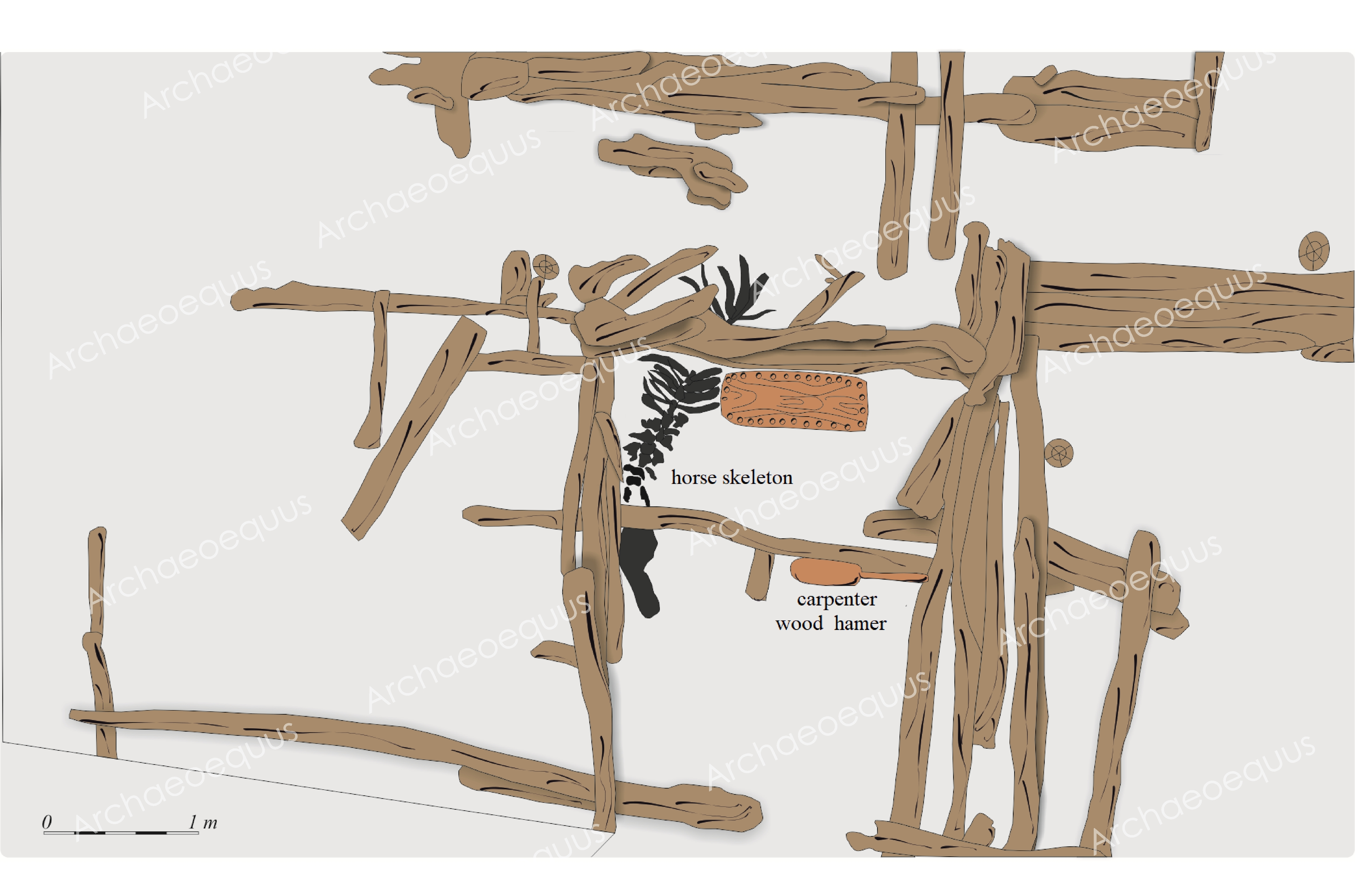
Gdańsk, site 1 (Na Dylach St.). Skeletal deposit of horse under wooden structure of rampart No 3 (Archive
of Archaeological Museum in Gdańsk, redrawn by J. Sawicka).