Żółte
Żółte, stanowisko 33., Osada. Crannog. Moduł D.Inw. Nr 23/08, wykop 30/w/08.
Chronologia: 1. poł. XI–XI w./XII w.
Elementy przegubowe osobnika o wysokości w kłębie 140,8 cm.
Literatura: Chudziak i Kaźmierczak 2014; Makowiecki i Makowiecka 2014c.
|
Anatomical element |
NISP |
Descriptions and biometrical data |
|
Carpal bones |
3 |
right |
|
Third metacarpal bone |
1 |
right; bone IV fused to the diaphysis of bone III (Makowiecki & Makowiecka 2014c,Figure 7.29., p. 351); GL = 229.1: Bp = 51; SD = 31.8; Bd = 48.8; WH = 140.8 cm |
|
Second metacarpal bone |
1 |
right |
|
Fourth metacarpal bone |
1 |
right |
Żółte, stanowisko 33., Crannog. nr inw. Nr 58/06, wykop 25/w/06, depozyt podwodny,
Chronologia: 1. poł. XI–XI w./XII w.; 14C: 980 pz ± 35 pz; Poz-21867 (wg Chudziak i Kaźmierczak 2014), prawdopodobieństwo 68,20%: 1024AD (22,0%) 1048AD; 1082 r. (37,7%) 1130 r.; 1138 r. (8,6%) 1150 r.; prawdopodobieństwo 95,40%: 995AD (3,5%) 1006AD; 1016 r. (91,9%) 1158 r. n.e.
Szkielet samca w wieku 9-10 lat, wysokość w kłębie 131,2-137,7 cm (średnio 133,1 cm).
Literatura: Chudziak i Kaźmierczak 2014; Makowiecki & Makowiecka 2014c i ten projekt.
|
Anatomical element |
NISP |
Descriptions and biometrical data |
|
Skull |
1 |
whole, canines abraded to about 1/3 of their height, degree of incisor tooth wear about 9 years old; minor hook degenerations – small nodules on both lacrimal bones, also minor degenerations near the nuchal crest, pathological changes – small flat growths on the nasal and buccal bones on the right side, similar degenerations of a smaller scale on the left side; skull surface smooth without traces of anthropogenic; preserved; nuchal crest concave, minimal asymmetry to the left; occipital protuberance flat and smooth; frontals minimally concave; B-P = 471.7, WH = 134.9 cm, AP = 518.5, WH = 140.3 cm |
|
Mandible |
1 |
whole, left and right, slight abrasion of P2 edges; an anomaly in the clash of corners; canines preserved in their entirety without signs of abrasion |
|
Hyoid bone |
2 |
left and right |
|
Atlas |
1 |
degeneration of the area of muscle attachments on the dorsal arch, senile changes |
|
Axis |
1 |
|
|
Cervical vertebrae |
4 |
III–VI |
|
Thoracic vertebrae |
4 |
XV–XVIII |
|
Lumbar vertebrae |
1 |
I, from the lumbar vertebra II to the last four thoracic vertebrae, degenerative changes at the articular connections of the cranial and caudal processes of all vertebrae |
|
Lumbar vertebrae |
1 |
II, pathological changes on the right side of the diaphysis – accumulation of spongy substance – cyst |
|
Lumbar vertebrae |
4 |
III–VI |
|
Sacrum |
1 |
|
|
Ribs |
3 |
left, for VII changes in the form of deformation of the middle part of the rib, probably as a result of mechanical trauma, for XII changes in the form of thickening of the distal part due to a long-term inflammatory process, which could have been caused by mechanical trauma, for XIV changes in the proximal part in the form of thickening of such same character as XII |
|
Ribs |
15 |
right, no pathological changes |
|
Ribs |
9 |
cartilage |
|
Ribs |
12 |
left |
|
Scapula |
1 |
left, 123, ++, HS = 344.3; DHA = 328; SLC = 63.4; GLP = 91.1; LG = 53.6; BG = 47 |
|
Scapula |
1 |
right, 123; HS = 345; DHA = 332.6; SLC = 61.8; GLP = 90.4; LG = 55.8; BG = 46 |
|
Humerus |
1 |
left, 123, ++; traces of cuts near the distal epiphysis on the lateral side and in the vicinity of the epicondyle, analogous traces of cuts visible on the nodule of the ulna bone on the lateral side; GL = 278; Bp = 96.8; SD = 34.8; Bd = 77.5; BT = 71.8; WH = 131.2 cm |
|
Humerus |
1 |
right, 123, ++; GL = 278: Bp = 97.2; SD = 36.4; Bd = 77.9; BT = 72.4; WH = 131.2 cm |
|
Radius & ulna |
1 |
left, 123, ++; GL = 319.5; Bp = 80.5; SD = 39.1; Bd = 72.5; WH = 131.8 cm |
|
Radius & ulna |
1 |
right, 123, ++; GL = 321.2; Bp = 80; SD = 39.4; Bd = 74.1; WH = 132.5 cm |
|
Radial carpal bone |
2 |
left and right |
|
Central carpal bones |
2 |
left and right |
|
Ulnar carpal bone |
1 |
right |
|
Accessory carpal bone |
1 |
right |
|
Third carpal bone |
2 |
left and right |
|
Fourth carpal bone |
2 |
left and right |
|
Third metacarpal bone |
1 |
left, 123.++, bone II fused to III, IV – traces of fusion not preserved; GL = 223.4; Bp = 50.3; SD = 32.8; Bd = 49.6; WH = 137.7 cm |
|
Third metacarpal bone |
1 |
right, 123, ++, bone II and IV firmly fused to the diaphysis of bone III; GL = 222.3; Bp = 50; SD = 33.2; Bd = 49.5; WH = 137.1 cm |
|
Pelvis |
1 |
left and right, whole; pubic symphysis fused, in the caudal part from the ventral side visible signs of degeneration, male |
|
Femur |
1 |
left, 123, ++; GL = 380; Bp = 118.8; SD = 39.4; Bd = 94.6; WH = 132 cm |
|
Femur |
1 |
right 123, ++; GL = 379: Bp = 117.8; SD = 39.4; Bd = 91.7; WH = 131.6 cm |
|
Patella |
1 |
left |
|
Tibia |
1 |
left, 123, ++, GL = 335; Bp = 96.3; SD = 40.7; Bd = 73.3; WH = 132 cm |
|
Tibia |
1 |
right, 123, ++; GL = 335; Bp = 94.9; SD = 41.3; Bd = 72.1; WH = 132 cm |
|
Proximal phalanx ant. |
1 |
right, GL = 82; Bp = 54.6; SD = 35.3; Bd = 47.9; WH = 127.7 cm |
|
Middle phalanx ant. |
1 |
right; GL = 44.7; Bp = 52; SD = 44.7; Bd = 50.9; WH = 123.3 cm |
|
Middle phalanx ant. |
1 |
left; GL = 44.5; Bp = 52.7; SD = 45.1; Bd = 52.8; WH = 122.7 cm |
|
Sesamoid bones |
1 |
even |
|
∑ |
88 |
Żółte, stanowisko 33. Crannog. nr inw. Nr 72/05, wykop 26/w/07, złoże podwodne (ryc. 27).
Chronologia: początek XI–XI w./XII w.; 14C: 960 ± 50 lat p.z.; MKL-7012 (wg Chudziak i Kaźmierczak 2014), prawdopodobieństwo 68,20%: 1028AD (15,7%) 1054AD; 1075 r. (52,6%) 1156 r.; prawdopodobieństwo 95,40%: 994AD (92,7%) 1180AD; 1190 r. (2,8%) 1206 r. n.e.
Szkielet samca w wieku 8-9 lat, o wysokości w kłębie 137,4-140,8 cm (średnio 139,5 cm).
Literatura: Chudziak i Kaźmierczak 2014; Makowiecki & Makowiecka 2014c i ten projekt.
|
Anatomical element |
NISP |
Descriptions and biometrical data |
|
Mandible |
1 |
male about 8-9 years old; wear of the anterior parts of the crown surface on both teeth P2. |
|
Hyoid bone |
2 |
right, left |
|
Atlas |
1 |
pathological changes in the anterior part of the left wing resulting from a mechanical injury causing fractures |
|
Axis |
1 |
whole |
|
Cervical vertebrae |
5 |
preserved whole, ++ |
|
Thoracic vertebrae |
7 |
on the corpus of the fifth vertebra at the cranial epiphysis, an epiostoma; distal epiphyses–traces of fusion with the diaphysis, from vertebra X to XV growths on the ventral edge of the diaphysis located on the left side; the penultimate vertebra is missing |
|
Thoracic vertebrae |
10 |
|
|
Lumbar vertebrae |
4 |
the last vertebra is missing |
|
Sacrum |
1 |
whole |
|
Caudal vertebrae |
1 |
|
|
Ribs |
12 |
cartilage |
|
Ribs |
2 |
left, on one distal epiphysis, slight degeneration from the medial side |
|
Ribs |
10 |
right, on one distal epiphysis, slight degeneration from the medial side |
|
Ribs |
15 |
left |
|
Sternum |
4 |
|
|
Scapula |
1 |
right, wholly preserved; SLC = 66.1; GLP = 93.4; LG = 55.9; BG = 49.9 |
|
Scapula |
1 |
left, wholly preserved; SLC = 64.8; GLP = 92.9; LG = 55.4; BG = 49.3 |
|
Humerus |
1 |
left; GL = 293.5; Bp = 92.2; SD = 35.6; Bd = 78; WH = 137.4 cm |
|
Humerus |
1 |
right; GL = 295; Bp = 91.7; SD = 37.1; Bd = 80; WH = 138 cm |
|
Radius & ulna |
1 |
left; GL = 341.8; Bp = 81.5; SD = 39.1; Bd = 73.3; WH = 140.7 cm |
|
Radius & ulna |
1 |
right; GL = 342; Bp = 81.8; SD = 39.3; Bd = 73.7; WH = 140.8 cm |
|
Radial carpal bone |
2 |
left and right |
|
Central carpal bones |
1 |
right |
|
Ulnar carpal bone |
1 |
left |
|
Third carpal bone |
2 |
left and right |
|
Second metacarpal bone |
1 |
left |
|
Third metacarpal bone |
1 |
left; GL = 224; Bp = 50.1; SD = 33.8; Bd = 49.8; WH = 138 cm |
|
Third metacarpal bone |
1 |
right; GL = 224.5; Bp = 50.6; SD = 34.7; Bd = 49; WH = 138.3 cm |
|
Fourth metacarpal bone |
2 |
left and right |
|
Femur |
1 |
left, ++ ; GL = 402; Bp = 120.4; SD = 38.9; Bd = 92.3; WH = 140.8 cm |
|
Femur |
1 |
right, ++; GL = 402; Bp = 121.2; SD = 39.8; Bd = 94.3; WH = 140.8 cm |
|
Patella |
1 |
right; H = 68.7; B = 67.7 |
|
Patella |
1 |
left; H = 68.6; B = 68.1 |
|
Tibia |
1 |
left, ++; GL = 354.5; Bp = 95.9; SD = 40.8; Bd = 69.8; WH = 139.8 cm |
|
Tibia |
1 |
right, ++; GL = 354.5; Bp = 96.7; SD = 41.3; Bd = 70.9; WH = 139.8 cm |
|
Tarsal bones |
1 |
IV, left |
|
Calcaneus |
1 |
left; GL = 110.9; GB = 52.7 |
|
Calcaneus |
1 |
right; GL = 111.6; GB = 52.7 |
|
Talus |
1 |
left; GH = 59.6; GB = 63.5; BFd = 51.9 |
|
Talus |
1 |
right; GH = 59.5; GB = 64; BFd = 52.4 |
|
Third metatarsal bone |
1 |
right, bone spavin type changes–fused central bone and tarsal bone III, very extensive inflammation; GL = 267; SD = 32.2; Bd = 49.5; WH = 139.6 cm |
|
Third metatarsal bone |
1 |
left, bone spavin type pathological changes – fused central bone and tarsal bone III, very extensive inflammation; GL = 267.5; Bp = 50.7; SD = 32.5; Bd = 49.1; WH = 139.9 cm |
|
Fourth metatarsal bone |
1 |
right |
|
Proximal phalanx ant. |
1 |
right; GL = 86.4; Bp = 52.8; SD = 35.5; Bd = 47.6; WH = 134.5 cm |
|
Proximal phalanx ant. |
1 |
left, fused with finger member II – very strong changes in the distal epiphysis of this member and the proximal epiphysis and diaphysis of the coronal bone – extensive growths |
|
Proximal phalanx post. |
1 |
left; GL = 84.3; Bp = 55.8; SD = 34.9; Bd = 46.1; WH = 133.5 cm |
|
Proximal phalanx post. |
1 |
right; GL = 84; Bp = 53.9; SD = 33.9; Bd = 45.5; WH = 133.1 cm |
|
Middle phalanx ant. |
1 |
left, fused with the proximal finger member |
|
Middle phalanx ant. |
1 |
right; GL = 46.2; Bp = 52.6; SD = 45.9; Bd = 51.8; WH = 127.4 cm |
|
Middle phalanx post. |
1 |
left; GL = 48.4; Bp = 51.6; SD = 42.3; Bd = 49.2; WH = 128.5 cm |
|
Middle phalanx post. |
1 |
right; GL = 47.8; Bp = 51.4; SD = 42.8; Bd = 49.3; WH = 126.9 cm |
|
Distal phalanx ant. |
1 |
left, pathological changes–growths from the cranial side |
|
Distal phalanx ant. |
1 |
|
|
Distal phalanx post. |
2 |
left & right |
|
∑ |
119 |
Żółte, stanowisko 33. Crannog. nr inw. nr 32/06, na południe od wyspy.
Chronologia: 1. poł. XI–XI w./XII w.
Elementy szkieletowe trzech osobników: samicy powyżej 20 lat o wysokości w kłębie 116,8-124,3 cm (średnio 121,1 cm), samca w wieku 12 lat o wysokości w kłębie 132,2 -136,0 cm (średnio 133,9 cm) i samica w wieku około 7-10 lat.
Literatura: Chudziak i Kaźmierczak 2014; Makowiecki & Makowiecka 2014c i ten projekt.
|
Anatomical element |
NISP |
Descriptions and biometrical data |
|
The first individual |
||
|
Mandible |
1 |
left and right, male about 12 years old, P2 – traces of abrasion of the tooth crown in the mesial part; left: LP-M = 166.3 |
|
Scapula |
1 |
right, 123; SLC = 59.2; GLP = 93 |
|
Humerus |
1 |
left, 123, ++; GL = 285; Bp = 85.7; SD = 32.9; Bd = 76.3; WH = 134 cm |
|
Humerus |
1 |
right, 123, ++; GL = 282.5; Bp = 83; SD = 33.4; Bd = 79.4; WH = 133 cm |
|
Radius & ulna |
1 |
left, 123, ++; GL = 326.4; Bp = 79.1; SD = 36.7; Bd = 71.3; WH = 134.6 cm |
|
Radius & ulna |
1 |
right, 123, ++; GL = 323.3; Bp = 79.5; SD = 36.9; Bd = 71.8; WH = 133.3 cm |
|
Femur |
1 |
left, 123, ++; GL = 383; Bp = 110.7; SD = 38.7; Bd = 90.8; WH = 133.2 cm |
|
Femur |
1 |
right, 123, ++; GL = 382; Bp = 114.1; SD = 39.8; Bd = 91.2; WH = 132.8 cm |
|
Tibia |
1 |
right, 123, ++; GL = 345; Bp = 92.1; SD = 39.6; Bd = 68.2; WH = 136 cm |
|
Tibia |
1 |
left, 123, ++; GL = 343.5; Bp = 93.7; SD = 38.6; Bd = 68.5; WH = 135.4 cm |
|
The second individual |
||
|
Mandible |
1 |
right, incisors preserved, individual over 20 years old – based on buccal teeth, malocclusion – crown M3 worn off almost completely, probably female |
|
Scapula |
1 |
right, 12; SLC = 79.9; GLP = 55.6 |
|
Humerus |
1 |
right, 123, ++; GL = 250.5; Bp = 82.2; SD = 31.5; Bd = 71.7; WH = 120.2 cm |
|
Humerus |
1 |
left, 123, ++; slight deformation; GL = 257; SD = 34.8; Bd = 69; WH = 122.8 cm |
|
Radius & ulna |
1 |
left, 123, ++; GL = 300.7; Bp = 71.8; SD = 30.4; Bd = 63.8; WH = 124.3 cm |
|
Pelvis |
1 |
right, wholly preserved, pubic symphysis not completely fused |
|
Pelvis |
1 |
left, wholly preserved; LA = 55.5 |
|
Femur |
1 |
left, 123, ++; heavily covered with organic remains; GL = 342; Bp = 98.4; Bd = 80; WH = 116.8 cm |
|
Tibia |
1 |
left, 123, ++; heavily covered with organic remains; GL = 308; SD = 35.8; Bd = 62.3; WH = 121.2 cm |
|
The third individual |
||
|
Cervical vertebrae |
1 |
V |
|
Cervical vertebrae |
2 |
VI-VII |
|
Thoracic vertebrae |
1 |
I |
|
Thoracic vertebrae |
3 |
from the withers region |
|
Thoracic vertebrae |
1 |
one of the last |
|
Lumbar vertebrae |
2 |
|
|
Lumbar vertebrae |
1 |
penultimate |
|
Lumbar vertebrae |
1 |
the last one, about 7-10 years (+/+-) |
|
Ribs |
7 |
right, preserved almost whole, between VII-XIII-XIV |
|
Ribs |
22 |
|
|
Scapula |
1 |
right, 12; SLC = 56.9 |
|
∑ |
61 |
|
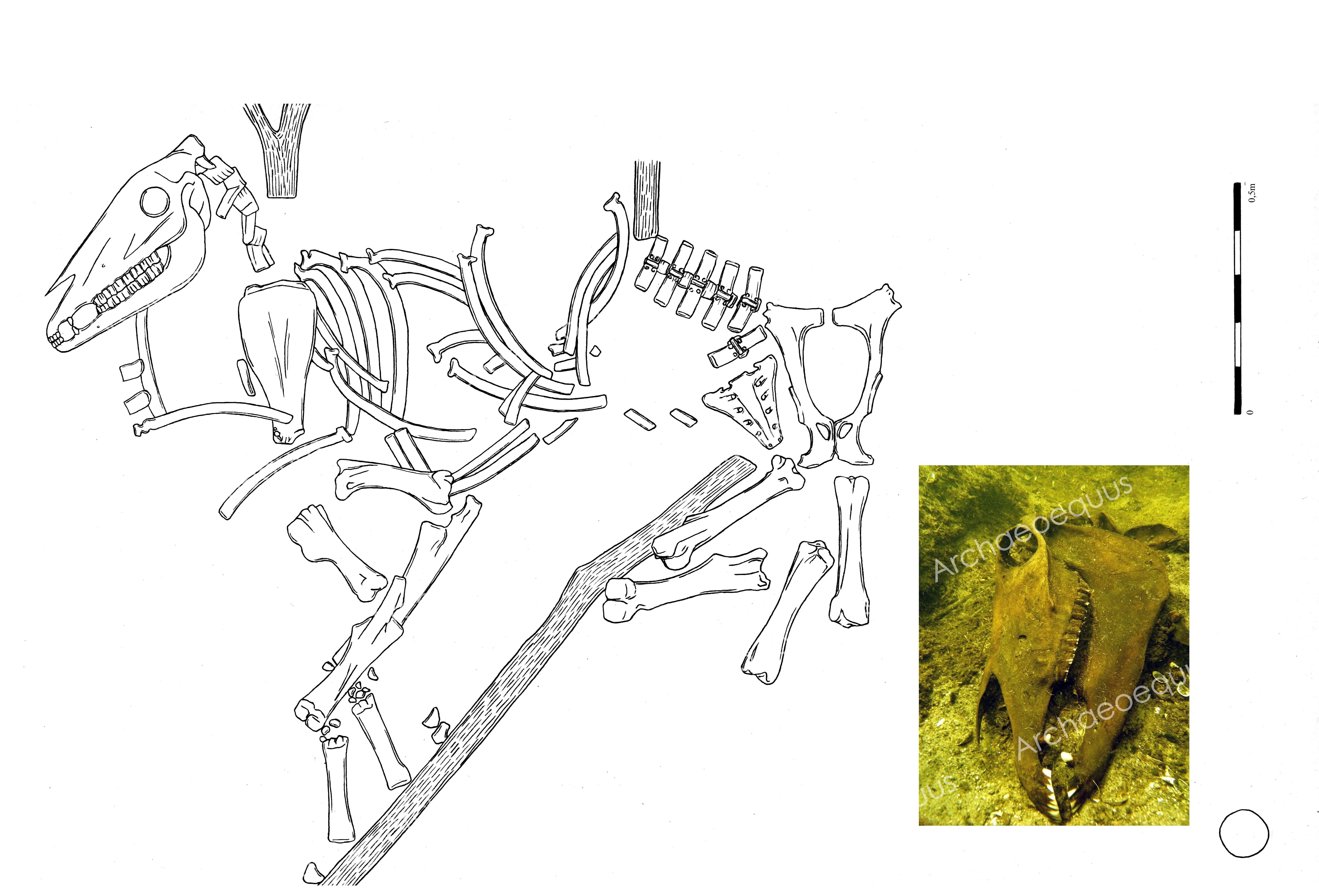
Żółte, site 33. Underwater skeletal deposit of horse, Inv. No. 58/06, trench 25/w/06 (after D. Makowiecki & M. Makowiecka 2014, drawing B. Kowalewska)
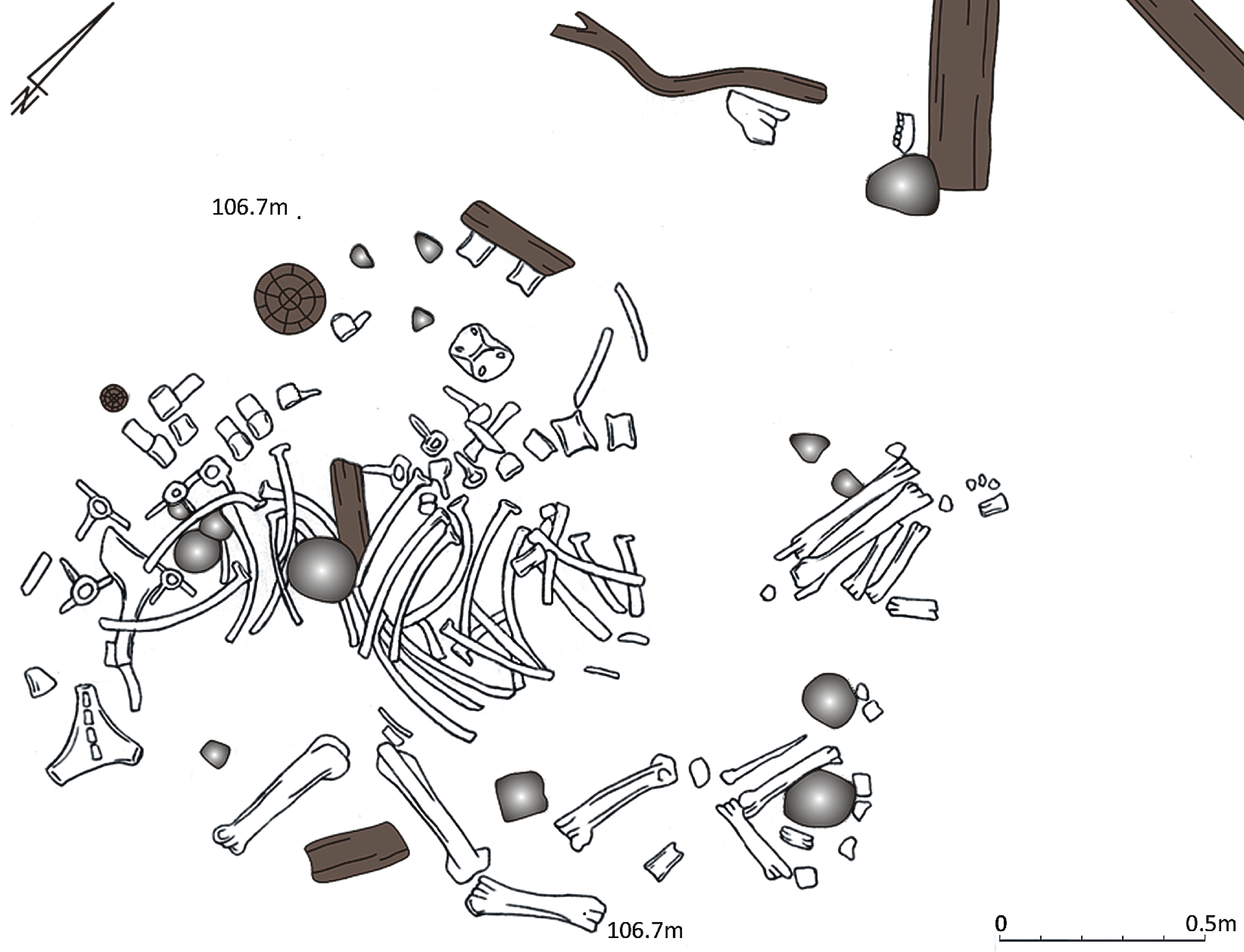
Żółte, site 33. Underwater skeletal deposit of horse, Inv. No. 72/05, trench 26/w/07 (after D. Makowiecki & M. Makowiecka 2014, drawing B. Kowalewska).